 Movie
Movie Controller
 Structure viewers
 About EMN search


-Search query
-Search result
Showing all 44 items for (author: prazak & v)
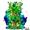
EMDB-18482: 
Herpes simplex virus 1 capsid (WT) vertices in perinuclear NEC-coated vesicles determined in situ
Method: subtomogram averaging / : Mironova Y, Prazak V, Vasishtan D
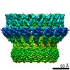
EMDB-18484:
Herpes simplex virus 1 nuclear egress complex (WT) determined in situ from perinuclear vesicles
Method: subtomogram averaging / : Mironova Y, Prazak V, Vasishtan D
EMDB-17974:
Pseudorabies virus cytosolic C-capsid (US3 KO) vertices determined in situ
Method: subtomogram averaging / : Prazak V, Grange M, Vasishtan D

EMDB-17975:
Pseudorabies virus primary enveloped (perinuclear) C-capsid (US3 KO) vertices determined in situ
Method: subtomogram averaging / : Prazak V, Grange M, Vasishtan D
EMDB-17976:
Pseudorabies nuclear C-capsids (US3 KO) vertices determined in situ
Method: subtomogram averaging / : Prazak V, Grange M, Vasishtan D

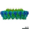
EMDB-18473:
Subtomogram average of pseudorabies virus nuclear egress complex helical form (UL31/34) determined in situ
Method: subtomogram averaging / : Prazak V, Grange M, Vasishtan D

EMDB-18474:
Subtomogram average of pseudorabies virus nuclear egress complex (UL31/34) determined in situ
Method: subtomogram averaging / : Prazak V, Grange M, Vasishtan D

EMDB-18479:
Pseudorabies virus cytosolic C-capsid (WT) vertices determined in situ
Method: subtomogram averaging / : Mironova Y, Prazak V, Vasishtan D
EMDB-18480:
Pseudorabies virus nuclear C-capsid (WT) vertices determined in situ
Method: subtomogram averaging / : Mironova Y, Prazak V, Vasishtan D
EMDB-18481:
Herpes simplex virus 1 cytosolic C-capsid (WT) vertices determined in situ
Method: subtomogram averaging / : Mironova Y, Prazak V, Vasishtan D
EMDB-18483:
Herpes simplex virus 1 nuclear C-capsid (WT) vertices determined in situ
Method: subtomogram averaging / : Mironova Y, Prazak V, Vasishtan D
EMDB-17704:
Subtomogram average of Vaccinia A10 trimer with open center from in vitro cores
Method: subtomogram averaging / : Turonova B, Liu J
EMDB-17708:
Subtomogram average of Vaccinia A10 trimer with tight center from in vitro cores
Method: subtomogram averaging / : Turonova B, Liu J
EMDB-17753:
Subtomogram average of Vaccinia A10 trimer from in situ cores
Method: subtomogram averaging / : Turonova B, Liu J
EMDB-15532:
Plasmodium falciparum sporozoite subpellicular microtubule with interrupted luminal helix determined in situ
Method: subtomogram averaging / : Prazak V, Ferreira JL, Vasishtan D, Grunewald K, Siggel M, Hentzschel F, Pietsch E, Kosinski J, Frischknecht F, Gilberger TW
EMDB-15534:
Plasmodium falciparum gametocyte subpellicular microtubule with 13 protofilaments determined in situ
Method: subtomogram averaging / : Ferreira JL, Prazak V, Vasishtan D, Grunewald K, Siggel M, Hentzschel F, Pietsch E, Kosinski J, Frischknecht F, Gilberger TW
EMDB-15535:
Plasmodium falciparum gametocyte subpellicular microtubule with 14 protofilaments determined in situ
Method: subtomogram averaging / : Ferreira JL, Prazak V, Vasishtan D, Grunewald K, Siggel M, Hentzschel F, Pietsch E, Kosinski J, Frischknecht F, Gilberger TW
EMDB-15536:
Plasmodium falciparum gametocyte subpellicular microtubule with 15 protofilaments determined in situ
Method: subtomogram averaging / : Ferreira JL, Prazak V, Vasishtan D, Grunewald K, Siggel M, Hentzschel F, Pietsch E, Kosinski J, Frischknecht F, Gilberger TW
EMDB-15537:
Plasmodium falciparum gametocyte subpellicular microtubule with 16 protofilaments determined in situ
Method: subtomogram averaging / : Ferreira JL, Prazak V, Vasishtan D, Grunewald K, Siggel M, Hentzschel F, Pietsch E, Kosinski J, Frischknecht F, Gilberger TW
EMDB-15538:
Plasmodium falciparum gametocyte subpellicular microtubule with 17 protofilaments determined in situ
Method: subtomogram averaging / : Ferreira JL, Prazak V, Vasishtan D, Grunewald K, Siggel M, Hentzschel F, Pietsch E, Kosinski J, Frischknecht F, Gilberger TW
EMDB-15539:
Plasmodium falciparum gametocyte subpellicular microtubule with 18 protofilaments determined in situ
Method: subtomogram averaging / : Ferreira JL, Prazak V, Vasishtan D, Grunewald K, Siggel M, Hentzschel F, Pietsch E, Kosinski J, Frischknecht F, Gilberger TW

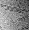
EMDB-15627:
Tomogram of the Mimivirus genomic fiber
Method: electron tomography / : Villalta A, Schmitt A, Estrozi L, Quemin ERK, Alempic JM, Lartigue A, Prazak V, Belmudes L, Vasishtan D, Colmant AMG, Honore FA, Coute Y, Gruenewald K, Abergel C
EMDB-15628:
Tomogram of the Mimivirus genomic fiber
Method: electron tomography / : Villalta A, Schmitt A, Estrozi L, Quemin ERK, Alempic JM, Lartigue A, Prazak V, Belmudes L, Vasishtan D, Colmant AMG, Honore FA, Coute Y, Gruenewald K, Abergel C

EMDB-15629:
Tomogram of the Mimivirus genomic fiber
Method: electron tomography / : Villalta A, Schmitt A, Estrozi L, Quemin ERK, Alempic JM, Lartigue A, Prazak V, Belmudes L, Vasishtan D, Colmant AMG, Honore FA, Coute Y, Gruenewald K, Abergel C

EMDB-15630:
Tomogram of the Mimivirus genomic fiber
Method: electron tomography / : Villalta A, Schmitt A, Estrozi L, Quemin ERK, Alempic JM, Lartigue A, Prazak V, Belmudes L, Vasishtan D, Colmant AMG, Honore FA, Coute Y, Gruenewald K, Abergel C
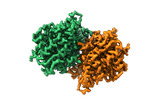
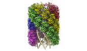
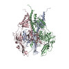
EMDB-13641:
Structure of the Mimivirus genomic fibre asymmetric unit
Method: single particle / : Villalta A, Schmitt A, Estrozi LF, Quemin ERJ, Alempic JM, Lartigue A, Prazak V, Belmudes L, Vasishtan D, Colmant AMG, Honore FA, Coute Y, Grunewald K, Abergel C
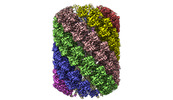
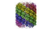
EMDB-14353:
Structure of the Mimivirus genomic fibre in its compact 6-start helix form
Method: helical / : Villalta A, Schmitt A
EMDB-14354:
Structure of the Mimivirus genomic fibre in its compact 5-start helix form
Method: helical / : Villalta A, Schmitt A, Estrozi LF, Quemin ERJ, Alempic JM, Lartigue A, Prazak V, Belmudes L, Vasishtan D, Colmant AMG, Honore FA, Coute Y, Grunewald K, Abergel C
EMDB-14355:
Structure of the Mimivirus genomic fibre in its relaxed 5-start helix form
Method: helical / : Villalta A, Schmitt A
PDB-7ptv:
Structure of the Mimivirus genomic fibre asymmetric unit
Method: single particle / : Villalta A, Schmitt A, Estrozi LF, Quemin ERJ, Alempic JM, Lartigue A, Prazak V, Belmudes L, Vasishtan D, Colmant AMG, Honore FA, Coute Y, Grunewald K, Abergel C
PDB-7yx3:
Structure of the Mimivirus genomic fibre in its compact 6-start helix form
Method: helical / : Villalta A, Schmitt A, Estrozi LF, Quemin ERJ, Alempic JM, Lartigue A, Prazak V, Belmudes L, Vasishtan D, Colmant AMG, Honore FA, Coute Y, Grunewald K, Abergel C
PDB-7yx4:
Structure of the Mimivirus genomic fibre in its compact 5-start helix form
Method: helical / : Villalta A, Schmitt A, Estrozi LF, Quemin ERJ, Alempic JM, Lartigue A, Prazak V, Belmudes L, Vasishtan D, Colmant AMG, Honore FA, Coute Y, Grunewald K, Abergel C
PDB-7yx5:
Structure of the Mimivirus genomic fibre in its relaxed 5-start helix form
Method: helical / : Villalta A, Schmitt A, Estrozi LF, Quemin ERJ, Alempic JM, Lartigue A, Prazak V, Belmudes L, Vasishtan D, Colmant AMG, Honore FA, Coute Y, Grunewald K, Abergel C
EMDB-12188:
Structure of DNA origami signpost from cryo-ET and subvolume averaging
Method: subtomogram averaging / : Silvester E, Vollmer B
PDB-7bho:
DNA origami signpost designed model
Method: subtomogram averaging / : Silvester E, Vollmer B, Prazak V, Vasishtan D, Machala EA, Whittle C, Black S, Bath J, Turberfield AJ, Gruenewald K, Baker LA
EMDB-11123:
Pre-fusion conformation of glycoprotein B of Herpes simplex virus 1
Method: subtomogram averaging / : Vollmer B, Prazak V, Vasishtan D, Jefferys EE, Hernandez-Duran A, Vallbracht M, Klupp B, Mettenleiter TC, Backovic M, Rey FA, Topf M, Gruenewald K
PDB-6z9m:
Pseudoatomic model of the pre-fusion conformation of glycoprotein B of Herpes simplex virus 1
Method: subtomogram averaging / : Vollmer B, Prazak V, Vasishtan D, Jefferys EE, Hernandez-Duran A, Vallbracht M, Klupp B, Mettenleiter TC, Backovic M, Rey FA, Topf M, Gruenewald K
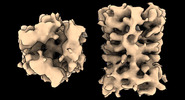
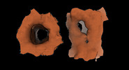
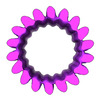
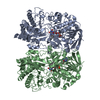
EMDB-10134:
CryoEM structure of murine perforin-2 ectodomain in a pre-pore form
Method: single particle / : Ni T, Yu X, Gilbert RJC
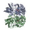
EMDB-10135:
CryoEM structure of murine perforin-2 ectodomain in a pore form
Method: single particle / : Ni T, Yu X, Gilbert RJC
EMDB-4995:
Subtomogram average of a part of the rabies lyssavirus ribonucleoprotein
Method: subtomogram averaging / : Riedel C, Vasishtan D, Prazak V, Ghanem A, Conzelmann KK, Ruemenapf T
EMDB-4471:
Cryo-SOFI enabling low-dose super-resolution correlative light and electron cryo-microscopy
Method: electron tomography / : Moser F, Prazak V, Mordhorst V, Andrade AM, Baker LA, Hagen C, Grunewald K, Kaufmann R
EMDB-4472:
Cryo-SOFI enabling low-dose super-resolution correlative light and electron cryo-microscopy
Method: electron tomography / : Moser F, Prazak V, Mordhorst V, Andrade AM, Baker LA, Hagen C, Grunewald K, Kaufmann R
EMDB-4473:
Cryo-SOFI enabling low-dose super-resolution correlative light and electron cryo-microscopy
Method: electron tomography / : Moser F, Prazak V, Mordhorst V, Andrade AM, Baker LA, Hagen C, Grunewald K, Kaufmann R
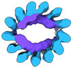
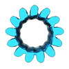
EMDB-3035:
The structure of YnaI implies structural and mechanistic conservation in the MscS-family of mechanosensitive channels
Method: single particle / : Bottcher B, Prazak V, Rasmussen A, Black SS, Rasmussen T
 wwPDB to switch to version 3 of the EMDB data model
